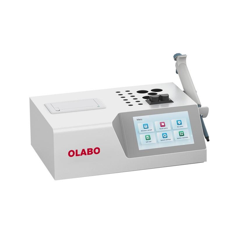
Hematology Analyzer
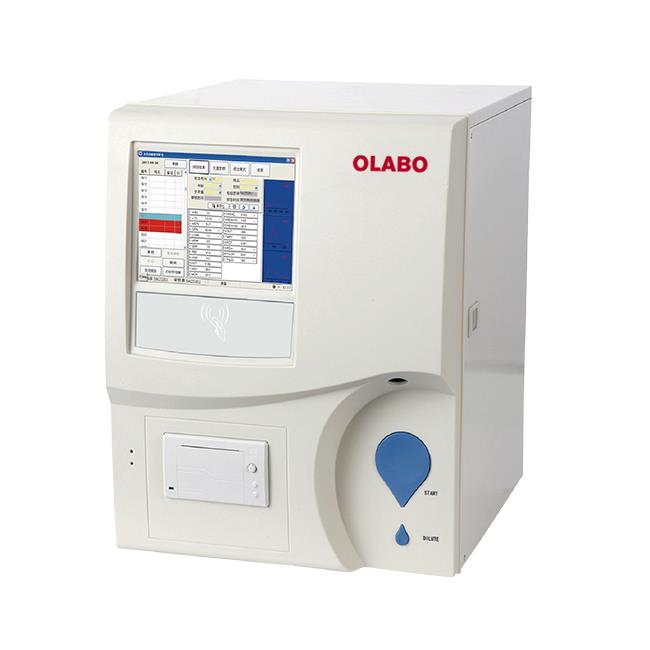
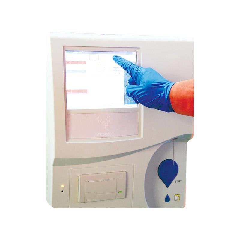
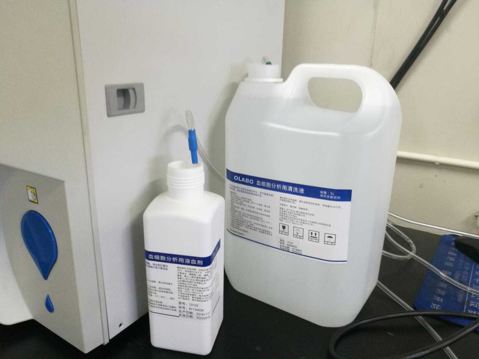
1.Brief Introduction
Hematology analyzers are also called hematology analyzers, hematology analyzers, hematology counters, etc. They are one of the most widely used instruments in hospital clinical testing. With the rapid development of computer technology in recent years, the technology of blood cell analysis has also changed from three groups. Turning to five groups, from two-dimensional space to three-dimensional space, and we have also noticed that many of the five classification technologies of modern blood cell analyzers use the same technologies as today’s very advanced flow cytometers, such as scattered light detection technology and sheath flow. Technology, laser technology, etc. This article focuses on the detection methods and applications of the quintile blood cell analysis instrument.
a.Development History
At the beginning of the 20th century, Moldland used a photoelectric device to count blood cells; in 1947, Lagerkranz used high-efficiency photomultiplier tubes, photoelectric scanning technology and dark-field illumination to detect and analyze blood cells, overcoming the Moldland photoelectric method. The problems that exist in the test can be used in clinical trials; in 1958, on the basis of predecessors, Kurt used the method of combining resistivity changes and electronic technology to develop a blood analyzer with relatively stable performance and convenient operation. It is a Kurt electronic blood cell counter. In the 1960s, various types of hematology analyzers based on Coulter's principle emerged and were widely used, gradually replacing traditional microscope operations. In the 1970s, various blood analyzers based on the laser sheath flow technology were developed on the Kurt principle. In the early 1980s, a dual-channel instrument was introduced. In addition to directly counting platelets, 14 parameters such as the total number and percentage of lymphocytes can also be obtained. Since the 1990s, some scholars have classified cells based on the structural differences between immature cells and mature cell membranes, especially the detection of immature cells, creating a new field for blood cell counters.
b.Fundamental
According to the different ways of acquiring blood cell signals, the principles can be summarized into five types: photoelectric, capacitive, resistive, centrifugal and laser scattering.
2.Detection method
a.Volume, conductivity, laser scattering method (VCS)
This is the classic analysis method used by the blood cell analyzer produced by Beckman-Coulter. It integrates three physical detection techniques and performs multi-parameter analysis on the cells in their natural primitive state. This method is also called volume, conductivity, and laser light scattering blood cell analysis. This technique uses a red blood cell hemolytic agent to be added to the specimen to dissolve the red blood cells, and then a stabilizer is added to neutralize the effect of the red blood cell lysis agent, so that the white blood cell surface, cytoplasm and cell volume remain stable. Then, the sheath flow technology is used to advance the cells into the Flowcell, and they are tested by the three technologies of the instrument VCS.
V represents the volume measurement method, which uses the classic Kurt patented technology to accurately analyze the cell volume with low-frequency current. Volume is an important parameter for distinguishing white blood cell subgroups. It can effectively distinguish lymphocytes and monocytes with significant differences in size.
C stands for high-frequency conductivity (Conductivity), which uses the principle of high-frequency electromagnetic probes to measure the differences in the internal structure of cells, which is also the company's patented technology. The cell membrane is conductive to high-frequency current. When the current passes through the cell, the chemical composition of the nucleus can change the conductivity of the current, and the amount of change can reflect the information of the cell contents. This parameter can be used to distinguish cell populations of similar size but different internal properties, such as lymphocytes and basophils, which differ in conductivity parameters due to their different nuclear properties.
S stands for laser light scattering (Scatter) measurement technology, which uses a monochromatic laser from a helium-neon laser source to scan each cell, and collects the scattered light (MALS) signal of the cell at an angle of 10° to 70°. The laser beam can penetrate cells, detect the condition of the cell core and the particles in the cytoplasm, provide information about the granularity of the cells, and can distinguish cell populations with different granular characteristics. For example, the scattered light of coarse particles in cells is stronger than that of fine particles, so it can be used to distinguish three types of granulocytes: neutrophil, eosinophil and basophil.
b.Joint detection technology of electrical impedance, radio frequency and cytochemistry
Typical models are SysmexSE-9000/SE-9500/XE-2100, etc. There are four different detection systems in this type of instrument. After the specimens are processed with special cell staining techniques, RF and DC techniques are used to classify and count white blood cells. It uses the following four detection systems:
Eosinophil detection system: This system uses electrical impedance to count. After the blood is separated by the blood separator, part of the blood is mixed with an eosinophil count hemolytic agent. The specific hemolytic agent dissolves or shrinks all cells except eosinophils. This liquid containing intact eosinophils passes through the impedance circuit count.
Basophil system: The detection principle of this system is the same as that of eosinophils, except that its hemolytic agent can only retain basophils in the blood.
Lymph, monocytes, granulocytes (neutrophils, eosinophils, basophils) detection system: The system uses electrical impedance and radio frequency combined detection methods, using a milder hemolytic agent, to nucleus and cells Type has little effect. There are two transmitters of direct current and high frequency on the inner and outer electrodes. Since direct current cannot reach the cytoplasm and nucleus, and radio frequency energy penetrates into the cell to measure the size of the nucleus and the number of particles, the number and level of these two different pulse signals comprehensively reflect the number of cells, size (DC), and nuclei and particles Density (RF). Because the size, cytoplasm content, nuclear shape and density of lymphocytes, monocytes and granulocytes are quite different, the ratio can be obtained by scanning.
Naive cell detection system: This system also uses electrical impedance to count. The principle is that the lipids on naive cells are less than those on mature cells. After adding sulfurized amino acids to the cell suspension, due to the difference in lipid occupancy, there are more sulfurized amino acids bound to naive cells than mature cells, and it is effective for hemolytic agents. Resistance, so it can keep the morphology of immature cells intact and lyse mature cells, which can be detected by impedance method.
c.Laser scattering and cytochemical staining technology
In the classification of white blood cells, the instrument uses two channels, one is the detection channel for peroxidase, and the other is the detection channel for basophils.
Peroxidase reaction (peroxidase, POX) is a common cytochemical staining method for blood smear staining, used to distinguish primitive cells from mature granulocytes, and to distinguish granulocytes from non-granulocytes. No blue-black particles appear in the stained cells as a negative reaction, small particles or sparsely distributed black particles are a weak positive reaction, and black coarse and dense particles are a strong positive reaction. Peroxidase is mainly present in granulocyte and monocyte cell lines. The reaction of various types of white blood cells to peroxidase is as follows: the early primordial granulocytes are negative, and the subsequent stages of promyelocytic contain peroxide. As the cell’s mature peroxidase content gradually increases, neutrophil granulocytes will have a strong positive reaction, eosinophils have the strongest peroxidase reaction, and basophils do not contain this The enzyme reaction was negative. In the monocyte system, in addition to the early primitive stage, naive monocytes and monocytes will have a weaker peroxidase reaction. Lymphocytes, naive red blood cells, megakaryocytes, etc. are all peroxidase negative reactions. The peroxidase detection channel is designed according to this principle. It detects every white blood cell passing through the flow counting cell, and the absorption rate of the peroxidase scattered light generated by laser irradiation. Of course, the reagents and auxiliary reagents have been improved.
1) Eosinophils with the strongest peroxidase positive;
2) Neutrophils with strong peroxidase positive nucleus granulocytes;
3) Larger monocytes with weak peroxidase positive;
4) Small, peroxidase-negative lymphocytes;
5) Unstained large cells that are larger than lymphocytes and are peroxidase-negative. The increase of such cells indicates that various types of naive or primitive cells may appear.
The detection principle used in the basophil channel is: a special basophil reagent removes the cell membrane of white blood cells except basophils, makes them naked and becomes smaller in size, and only keeps the basophils in their original state , The volume is significantly larger than other types of white blood cells.
d.Multi-angle polarized laser scattering technology
The blood cell analyzer launched by the American company ABBOTT uses a unique multi-angle polarized light scattering (MAPSS) technology in the classification of white blood cells. The blood cell counters produced by it are from CELL-DYN 3000, 3200, 3500, 3700, 4000, As well as Sapphire (sapphire), MAPSS technology is used in the classification of white blood cells. The basic principle of this technology is that cells produce scattered light at multiple angles under the irradiation of a laser beam. The four detectors of the instrument at four angles will receive the corresponding scattered light signals and then analyze and process them by a microprocessor. Place all kinds of cells in the corresponding positions on the scatter diagram, and calculate the white blood cell classification results.
The principle of Multi-Angle Polatised Scatter Separation of white cell (MAPSS) is that a certain volume of whole blood sample is diluted with sheath fluid in an appropriate proportion. The internal structure of the white blood cells is similar to the natural state. Because the basophil granules have hygroscopic properties, the structure of basophils is slightly changed. The internal osmotic pressure of red blood cells is higher than the osmotic pressure of the sheath fluid. The hemoglobin in the red blood cells is freed from the cells, and the water in the sheath fluid enters the red blood cells. The structure of the cell membrane is still intact. The liquid is the same, so red blood cells do not interfere with white blood cell detection. Under the action of the sheath flow system, the sample is concentrated into a small stream with a diameter of 30μm. This stream arranges the diluted blood cells individually, and then irradiates the cells with a laser beam, and the laser light is scattered in all directions. appear.
1) 0° is forward angle scattered light, which can roughly measure the cell size;
2) 10° is narrow-angle scattered light, which can measure the relative characteristics of cell structure and complexity;
3) 90° vertical light scattering, mainly to measure the internal particles and cell components of cells.
4) 90° is depolarized light scattering. Based on the particle's characteristic of depolarizing the polarized laser at a vertical angle, eosinophils are separated from neutrophils and other cells.
5) After measuring and analyzing a single white blood cell at the same time from these four angles, the white blood cells can be divided into 5 types: eosinophils, neutrophils, basophils, lymphocytes and monocytes. ABBOTT’s five-classification method is very interesting. Instead of traditional volume quantification, quantitative quantification is used. The determination of 10,000 cells will stop every time the count is completed.
3.Classification structure
a.classification
⒈According to the degree of automation: semi-automatic blood cell analyzer, automatic blood cell analyzer and blood cell analysis workstation, blood cell analysis pipeline;
⒉According to the detection principle: capacitance type, electrical impedance type, laser type, photoelectric type, combined detection type, dry centrifugal layered type and non-invasive type;
⒊The level of white blood cells classified by the instrument is divided into two groups, three groups, five groups, five groups + reticulocyte analyzer.
b.basic structure
The structure of each type of blood cell analyzer is different. But most of them are composed of mechanical systems, electrical systems, blood cell detection systems, hemoglobin measurement systems, computers and keyboard control systems, etc., in different forms.
c.computer system
Although different types of blood cell analyzers have different structures, they all have mechanical devices (such as automatic injection needles, blood separators, diluters, mixers, quantitative devices, etc.) and vacuum pumps to complete sample extraction and dilution , Transfer, mix, and move the sample into the detection area of various parameters. In addition, the mechanical system also performs the functions of cleaning pipes and removing waste liquid.
d.Electrical system
Main power supply, voltage components, temperature control device, automatic vacuum pump electronic control system and automatic monitoring, fault alarm and elimination of instruments in the circuit.
e.Blood cell detection system
The detection technology used in domestic blood cell analyzers can be divided into two categories: electrical impedance detection and light scattering detection.
⑴Impedance detection technology: It is composed of signal generator, amplifier, discriminator, threshold regulator, detection counting system and automatic compensation device. This type is mainly used in two-class or three-class instruments.
⑵ Light scattering detection technology: mainly composed of laser light source, detection area device and detector.
⑶Laser source: Argon ion laser is mostly used to provide monochromatic light.
⑷ Monitoring area device: It is mainly composed of a sheath flow device to ensure that the cell suspension forms a single array of cell flow in the detection fluid flow.
⑸ Detector: The scattered light detector is a photodiode, which collects the scattered light signal generated by the cells after laser irradiation; the fluorescence detector is a photomultiplier tube, which is used to receive the fluorescence signal generated by the cells after the laser is irradiated by fluorescent dyeing.
This type of detection technology is mainly used in "five categories, five categories + net weave red" instruments.
f.Hemoglobin measurement system
It is composed of light source (generally 540nm wavelength), lens, filter, flow colorimetric cell and photoelectric sensor.
g.Computer and keyboard control system
Make the detection process faster and more convenient.


